
Реставрация обширной фрактуры коронки
В данной статье будет рассмотрена методика реставрации обширной фрактуры центральных резцов верхней челюсти, а также отдаленные результаты лечения.
Об особенностях лечения травмированного зуба на вебинаре 1.3 и 2.3 - восстановление зуба вследствие травмы онлайн-курса Онлайн-школа реставрации Сергея Радлинского. 1.1. - 1.3.: прямая реставрация.
Клинический случай: 29-летняя пациентка с травмой центральных резцов верхней челюсти (Рис. 1, 2).
В центральном резце верхней челюсти справа был диагностирован поддесневой перелом (Рис. 3). Пациентка собрала и привезла недостающие фрагменты зубов (Рис. 4). Отломанные части центрального резца справа зацементировали на композитный адгезивный цемент (Рис. 5), а поддесневая фрактура была удалена (Рис. 6). Затем выполнили остеотомию с последующей гингивопластикой с целью восстановления естественной ширины (Рис. 7). Резец слева был восстановлен композитным материалом, так как не удалось найти отломанную часть зуба (Рис. 5).
Было проведено эндодонтическое лечение центрального резца на верхней челюсти справа, и спустя несколько недель диагностировали некроз пульпы в центральном резце слева, поэтому также требовалось эндодонтическое лечение (Рис. 8). Клиническое наблюдение в течение 8 лет подтвердило качественную работу врача (Рис. 9,10). Изображения рентген снимков (Рис. 11.)
Спустя 12 лет после травмы пациентка обратилась в стоматологический кабинет, чтобы перекрыть вестибулярные поверхности зубов керамикой (Рис. 12). Она была впечатлена красивыми улыбками ее друзей, которым установили керамические виниры, поэтому хотела повысить эстетику собственной улыбки.
Были подготовлены воск и модель, чтобы выяснить предпочтения пациента и рассмотреть возможные варианты реставрации. Пациентке понравился новый дизайн улыбки, решили изготовить два керамических винира. Были взяты оттиски и изготовлены виниры из полевошпатной керамики. После тщательной примерки во время приема, проверки краевого прилегания, контактных пунктов и моделирования окончательного цвета с глицериновым гелем, пациентку попросили одобрить заключительный этап работы.
Затем следовала адгезивная обработка виниров (10% HF в течение 90 секунд, с последующим тщательным полосканием, нанесением силана и адгезива). Наложен раббердам (Рис.15) и зацементированы виниры (Рис.16). Финальная клиническая ситуация (Рис.17-18).
Проверка результатов спустя 12 месяцев, клиническая картина через 13 лет после травмы (Рис.19,22).
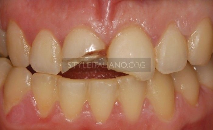
Рисунок 1. 29-летняя пациентка в стоматологическом кабинете сразу после травмы центральных резцов на верхней челюсти.
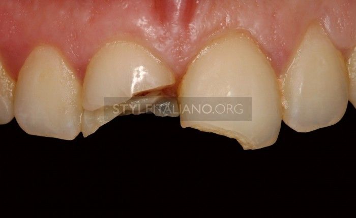
Рисунок 2. Фото травмированных зубов крупным планом.
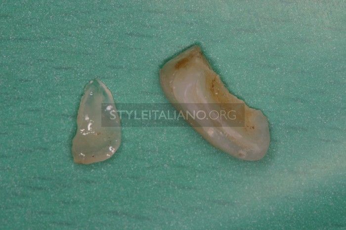
Рисунок 3. Отломанные фрагменты зуба.
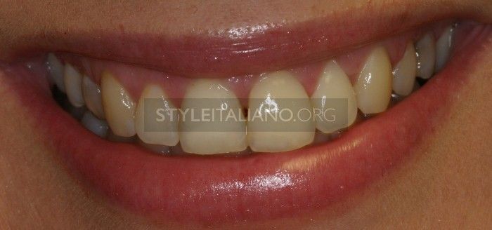
Рисунок 4. Клиническая картина после прикрепления отломанной части к 1.1, композитная реставрация 2.1.
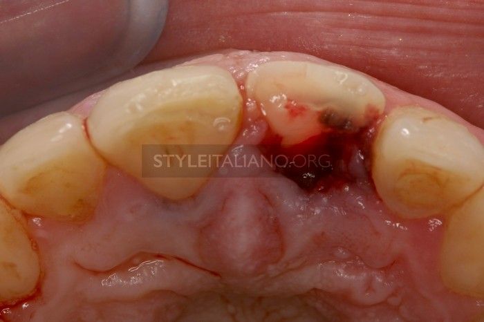
Рисунок 5. Поддесневая сломанная часть зуба была удалена.
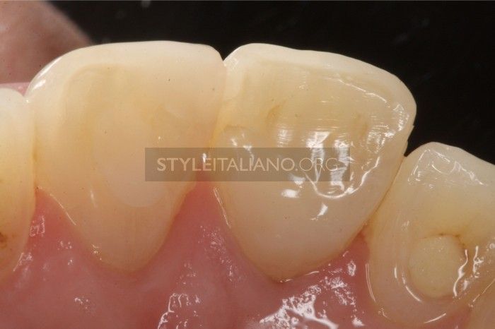
Рисунок 6. Остеотомия с последующей гингивопластикой с целью восстановления естественной ширины зуба.
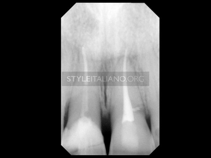
Рисунок 7. Зубы 1.1 и 2.1 после эндодонтического лечения.
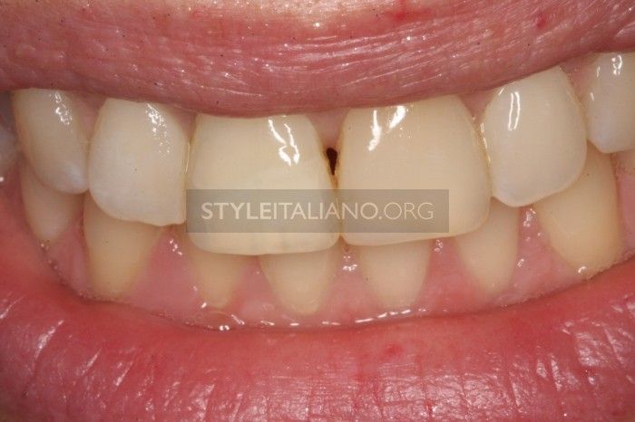
Рисунок 8. Клиническое наблюдение в течение 8 лет подтвердило качественно выполненную работу.
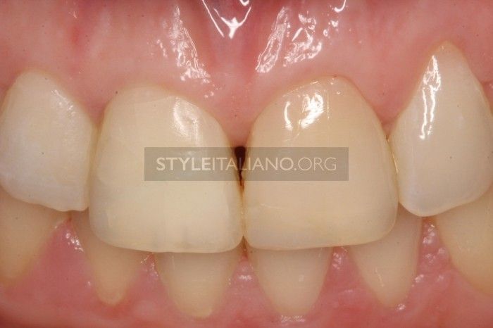
Рисунок 9. 1.1, 2.1 спустя 8 лет после травмы.
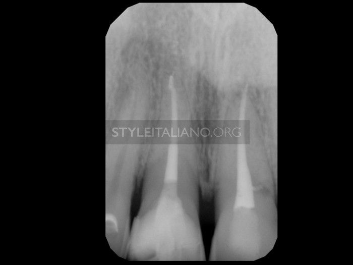
Рисунок 10. Рентген снимок зубов.
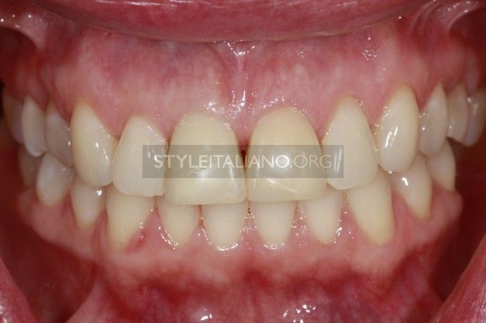
Рисунок 11. Спустя 12 лет пациентка обратилась в стоматологический кабинет с просьбой перекрыть центральные резцы керамическими винирами.
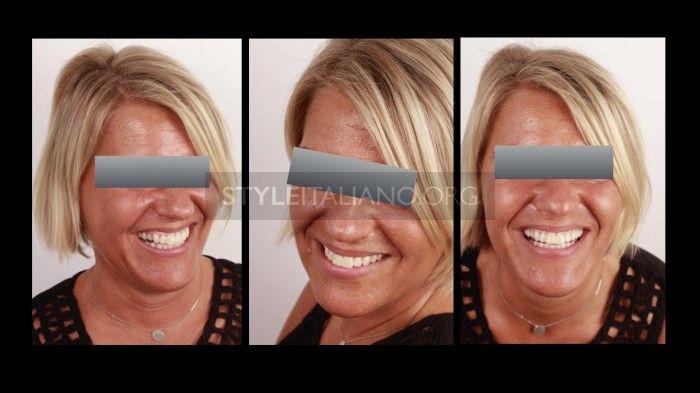
Рисунок 12. Была подготовлена модель, чтобы выяснить предпочтения пациента и рассмотреть возможные варианты реставрации.
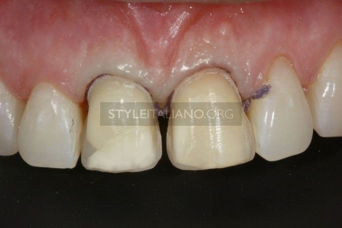
Рисунок 13. Отпрепарированные зубы под виниры.
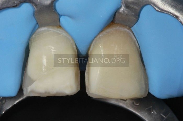
Рисунок 14. Изоляция перед цементировкой виниров.
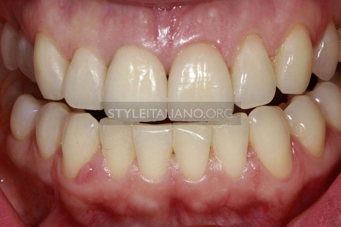
Рисунок 15. Клиническая ситуация после цементировки виниров на зубах (1.1,2.1).
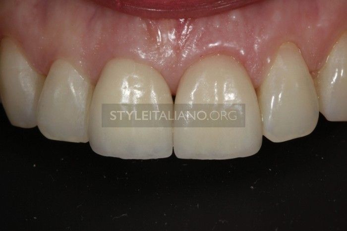
Рисунок 16. Фото реставраций крупным планом.
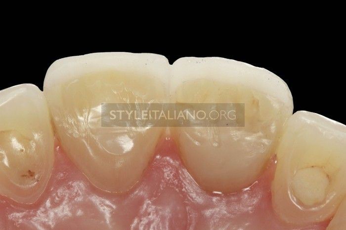
Рисунок 17. Вид реставраций с небной поверхности.
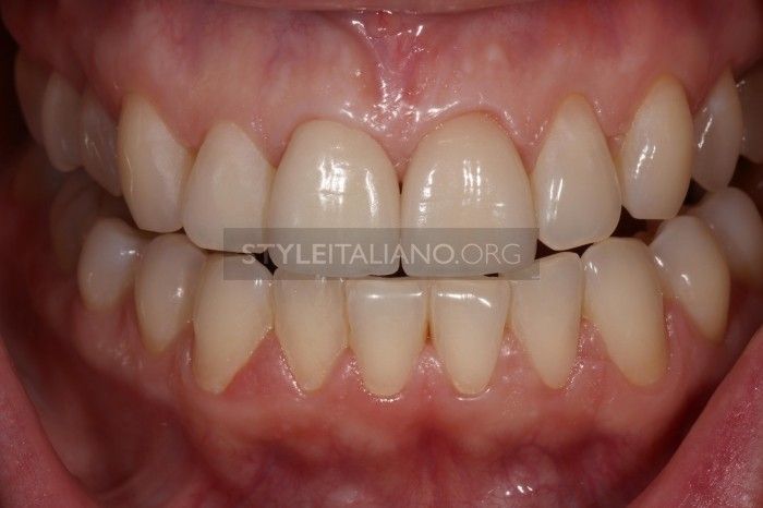
Рисунок 18. Контроль спустя 12 месяцев после цементировки.
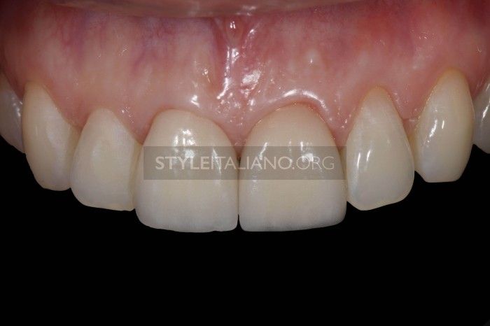
Рисунок 19. Прицельное фото 1.1 и 2.1 восстановлены винирами. 13 лет после травмы.
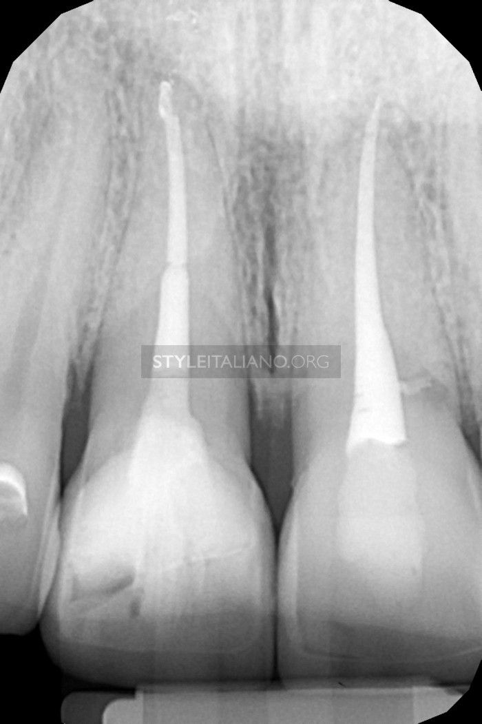
Рисунок 20. Рентген снимок 1.1, 2.1 один год после цементировки и 13 лет после травмы.
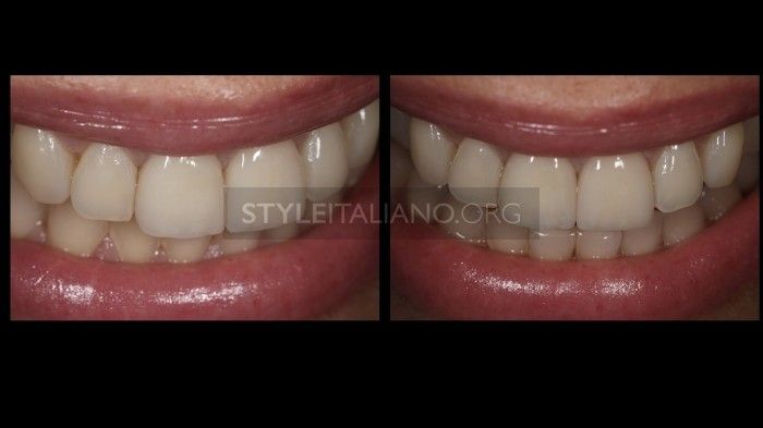
Рисунок 21. Улыбка счастливой пациентки спустя 13 лет после травмы центральных резцов верхней челюсти.
Выводы
- Минимально инвазивная адгезивная стоматология в отдельных случаях может быть успешной альтернативой лечению имплантатами при обширных переломах зубов.
- Необходимо учитывать тот факт, что каждый конкретный клинический случай индивидуален и требует тщательно составленного комплексного лечения.
Подробнее о композитных реставрациях на онлайн-курсе Концепция биомиметики: протоколы композитной реставрации.
http://www.styleitaliano.org/